Draft Summary of CPR guidelines
CPR – Cardio Pulmonary Resuscitation is the procedure done in patients with cardio/respiratory arrest to maintain ventilation and circulation to vital organs and to revive them back to life, using various maneuvres, drugs & equipment.
BLS – Basic Life Support comprises CPR done without any drugs or devices – just our hands, for chest compressions, and our mouth, for mouth-to-mouth breathing. Some guidelines also include the use of masks for mouth-to-mask breathing, an Ambu bag, and automated defibrillators in the BLS guideline.
ACLS – Advanced Cardiac Life Support – comprises the use of various drugs & devices in CPR – the defibrillator, endotrachel intubation, adrenaline, dopamine, atropine, temporary pacing, etc.
In whom is CPR done?
In a person who develops cardiac & / or respiratory arrest.
Often one of them will cease to function but, soon, in 2–3 minutes, the other will also stop functioning. So DO NOT Wait for the other organ also to stop working before starting CPR. The moment the heart or lungs stop working, begin CPR.
If the heart stops beating and there is no pulse, begin chest compressions.
If the lungs stop breathing properly / adequately, begin Ambu ventilations.
What are the causes of cardiac / respiratory arrest?
CRA is almost always the end event that precedes death of a person. Clinically, a person is declared dead on the basis of absent cardiac & respiratorty functioning. (as opposed to Brain Death which has different diagnostic criteria).
Cardiac arrest is commonly due to Coronary Artery Disease – like Acute Myocardial Infarction (which can cause VF), other heart diseases (valvular, cardiomyopathy, CHF, etc.), hypoxemia, other end stage systemic diseases, metabolic & electrolyte disturbances, poisonings & toxins, etc.
Respiratory arrest can be due to any disease from respiratory centre to the lungs, that depresses ventilation or prevents gaseous diffusion in the lungs. Causes, thus, can be head injury, strokes (CVA), poisonings / drugs causing CNS depression, foreign body / obstruction of airways, other lung diseases, COPD, etc.
EMD / PEA – (see below) is often due to causes like ( Hs & 4 Ts) –
Tamponade, Tension pneumothorax, Thrombosis (Pulmonary / Coronary), Toxin/ poisoning
Hypovolemia, Hypo/hyper kalemia, Hypothermia, Hypoxia, Hydrogen ion (acidosis/ alkalosis)
How to recognize cardiorespiratory arrest?
- The patient suddenly becomes unresponsive – a talking person suddenly stops, they seem to stare into nowhere, and are ‘cut off’ from the environment.
- More often, however, the patient seems to have a tonic spasm, almost like a seizure or a decerebrate rigidity. (we can differentiate because in ‘arrest’ there is no pulse or cardiac output).
- When we feel for the carotid pulse, there is none;
- Respirations have ceased or are abnormal in the form of gasping or grunting or labored, occasional breathing.
(as mentioned earlier, dont wait for both heart & lungs to stop working; as soon as one stops, begin appropriate resuscitative measures, getting ready to institute full CPR if needed).
Types of cardiac arrest (By ECG)
By ECG we can differentiate 3 types of cardiac arrest –
- VF (ventricular fibrillation)
- Asystole
- PEA / EMD – Pulseless Electrical Activity or Electromechanical Dissociation
Clinically all 3 cause same features – unresponsiveness, absent pulse & breathing.
It is important to differentiate the 3 – because treatment is different –
– for VF – defibrillate
– for asystole – pace
– for PEA/EMD – find out the cause & treat it
VF has a slightly better chance of successful resuscitation.
Steps of BLS
1. First recognise & confirm cardiorespiratory arrest by –
– pt. being unresponsive / having a tonic spasm of the body
– no carotid pulse
– no or abnormal breathing
(ECG may show VF or a flat line in VF or asystole).
2. Call for help / to get the defibrillator, etc.
3. Confirm the patient is on a firm / hard flat surface.
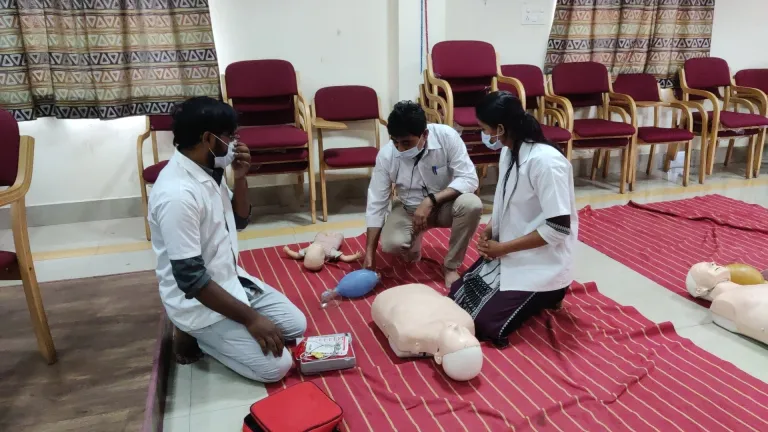
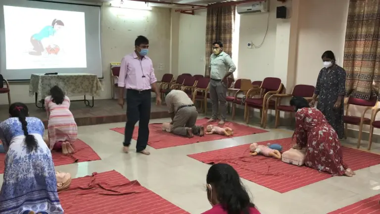
4. Begin chest compressions – Kneel on the bed and lock both hands’ fingers and place the heel of your hand on the lower half of the sternum on the midline. Compress the chest, with elbows straight, 5-6cms. – with your lumbosacral joint acting as fulcrum as you compress & release the chest. Compress at a rate of 100-120 min. (Time yourself) & count.
5. Every 30 compresssions, you or your colleague give 2 ventilations – if you are alone, give mouth to mask (or mouth to mouth) but not Ambu bag because it takes more time. If you are two, your colleague will give 2 breaths using an Ambu bag-mask.
6. Every 5 cycles (of if the defibrillator is getting ready to shock) or about 2 minutes, you can switch roles – you will give Ambu vetilations and your colleague will do chest compressions.
7. Do not interrupt CPR – unless the defibrillator is attached and is readying to shock or you think the patient is moving (signs of life). If the patient shows no signs of recovery (breathing/ movement), you may check for signs of life (pulse / breathing) once after 2-4 minutes of good CPR.
8. If an AED (Automated External Defibrillator) is available and has been connected, it is programmed to analyse the ECG every 2 min. or so. During the time it is analysing, you two must switch roles to avoid fatigue of the compression person.
9. The AED will analyse the patient’s ECG and
– if it is VF, it will advise a shock, it will charge the defibrillator and ask you to deliver the shock.
– if the rhythm is asystole, it will say there is no need of shock but continue CPR.
– if it recognises a rhythm (PEA /EMD) it will say there is no need for shock and ask you to do CPR if needed, or it may ask you to check for the pulse – if so, check pulse / breathing and act accordingly –
– if both are normal, the patient has recovered and you can shift the patient to the ICCU.
– if pulse is absent, continue CPR & search for a cause of PEA/EMD.
– if the pulse is present and breathing is abnormal/ absent, begin rescue breathing at a rate of 1 breath every 6 seconds.